Introduction
Do you want to know how your heart is doing?
Get it TESTED!!!
As Cardiologists, we play a crucial role in assessing and managing your cardiovascular health, a task that often involves ordering a variety of diagnostic tests to gain insights into your heart’s structure and function.
Your heart is a very complex structure, made up of a pump, valves, arteries and an electrical system. There is no one test you can do to check your heart out. It requires a number of diffent modalites to see if your heart if functioning as it should be.
These cardiovascular tests help in identifying potential issues, guiding treatment decisions, and monitoring progress.
In this big picture overview, we will explore some of the common heart tests that your cardiologist may order.
Electrocardiogram (ECG or EKG):
Let’s check your the basics of your hearts electrical system!
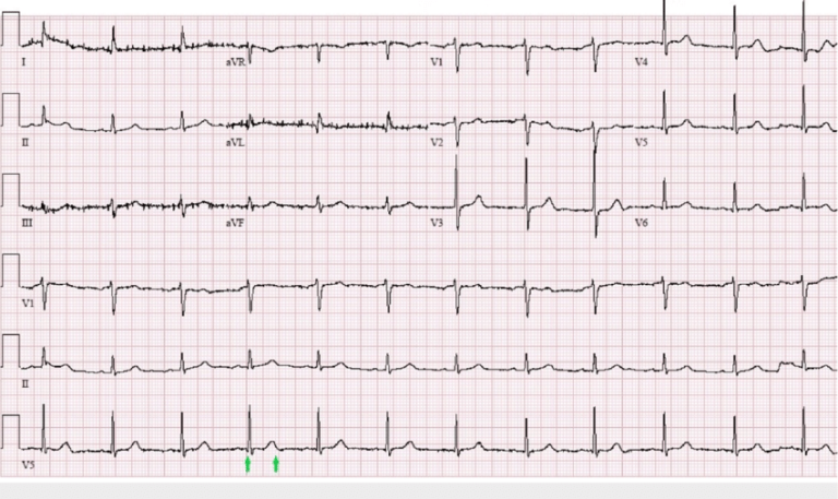
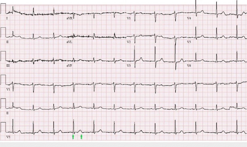
The EKG is our foundational test used to record the electrical activity of your heart over a short period of time.
Electrodes are placed on your skin to measure the heart’s rhythm and detect abnormalities such as arrhythmias, conduction abnormalities, and signs of ischemia. It’s a quick and non-invasive test that provides valuable information about the heart’s electrical function.
EVERYONE needs an EKG.
Echocardiogram:
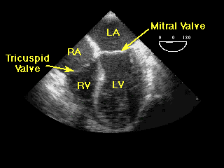
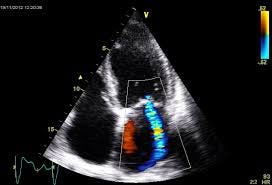
An echocardiogram (we usually just say “echo”) is an ultrasound of the heart that produces detailed images of the heart’s structure and function.
This test helps us assess the size and thickness of the heart chambers, the condition of heart valves, and the efficiency of the heart’s pumping function. It is particularly useful in diagnosing conditions such as heart failure, valvular heart disease, and congenital heart abnormalities. An echo does not see your heart arteries or your electrical system.
An echo is easy to do: you just lie on your side for about 45 minutes while a expert echo technian looks at your heart from a variety of views. There are no IVs and no prep is needed.
Echos are a great way to get a lot of good information about the health of your heart.
Your cardiologist may order an echo to evaluate:
- Heart murmurs
- Evidence of a prior heart attack
- Evidence that high blood pressure is affecting your heart
- Your heart valves structure and function
- Heart issues that you may have been born with (like a hole in your heart)
- and many, many other reasons…
Stress Tests: Treadmill Stress and Stress Echo
Stress tests, also known as treadmill tests or exercise tests, evaluate your heart’s response to physical activity. Stress testing requires your heart to be under “stress”.
An exercise stress test evaluates how well your heart handles work. As your body works harder during the test, it requires more oxygen, so the heart must pump more blood. The test can show if the blood supply is reduced in the arteries that supply the heart.
We do this by either having you walk on a treadmill or receive a medication that simulates the effects of exercise while their heart activity is monitored.
Stress tests are crucial in checking our your sympotms of chest pain or shortenss of breath and detecting coronary artery disease, assessing exercise tolerance, and evaluating the effectiveness of cardiac medications.
Exercise Treadmill Test (ETT):
Cardiologist call this a plain old “vanilla” stress test. It’s just you, a few EKG leads and the treadmill.
Most people who get into the hands of a cardiologist will probably not be getting a regular treadmill stress test. They are OK for a small subset of people but there are too many false positive and too many false negative results: this means that the test may give your cardiologist inaccuate info about your heart. The results are even more misleading for women than for men.
Stress Echocardiogram (Stress Echo):
A more accurate type of stress test is a stress echocardiogram- or stress echo.
This test is similar to a ETT but now we add an additional tool to the test to check your heart out. First, we do an echo of your heart while you are at “rest” before any type of exercise
Then, we hit you with exercise on a treadmill.
Now get ready for the challenging part: once you hit your maximum exercise on the treadmill, then we lie you down on your side right away and take a second set of echo images of your heart. This is a bit of a challege because our technician may ask you to hold your breat for a few seconds so she can get a great look at your pumping heart.
This test gives us three great ways to look at your heart: how you feel with exercise, we analyze your EKG and we look directly at your heart pump and how it responds to the stress of working out.
ZIO Patch or Holter Monitor:
We use an EKG in the office to take a quick snapshot of how your electrical system is functioning. Often times, however, we need to take a much longer look at your electrical system- your heart rhythm.
Many of our patients have symptoms such as palpitations, heart racing, heart skipping or thumping. They may have passed out or are feeling light headed at times.
These types of symptoms are concerning for abnormal heart rhythm, like atrial fibrillation. For this, we need a way to monitor your hearts rhythm for much longer periors- like a week or two.
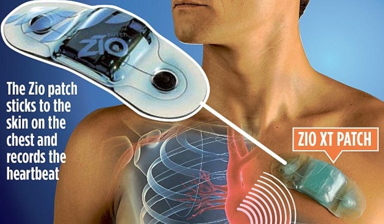
For continuous monitoring of the heart’s electrical activity over an extended period, we will prescribe a ZIO Patch. This portable device records the heart’s rhythm for one or two weeks, helping identify intermittent arrhythmias that may not be captured during a standard ECG.
We may put on the monitor right in the office or just mail it to you with great instructions on how to wear it and return it.
Cardiac CT Scan: A Coronary Artery Calcium Score or CCTA
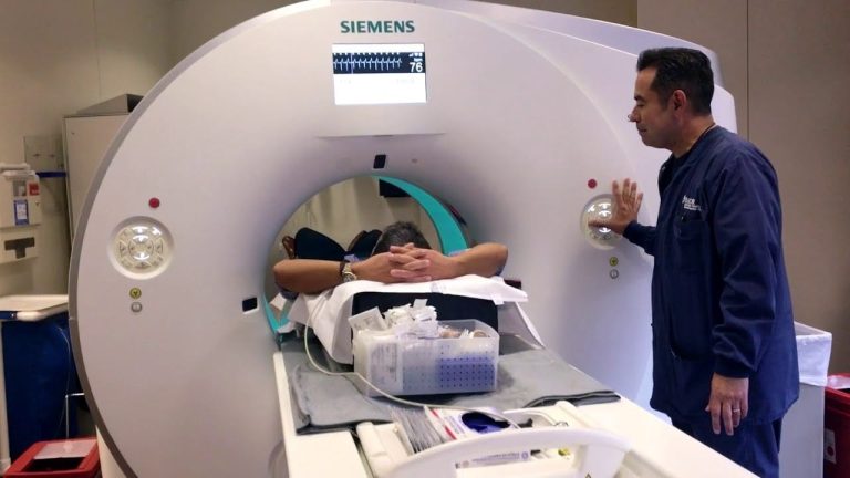
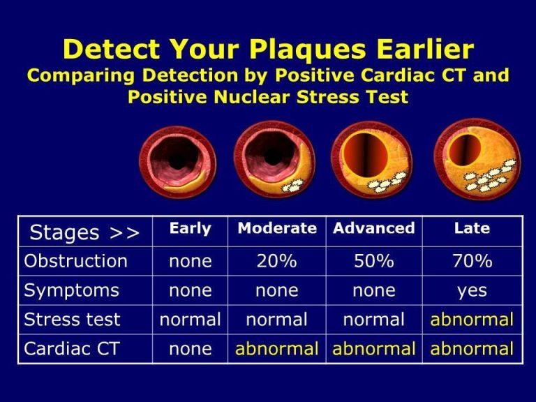
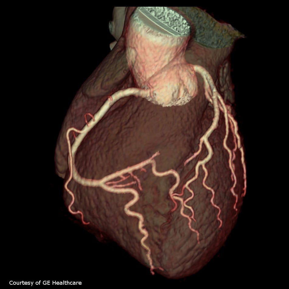
Cardiac computed tomography (CT) scans use X-rays to create detailed cross-sectional images of the heart and blood vessels. These amazing tests have come a long way in the last few years.
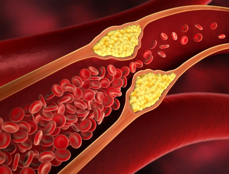
These test are valuable in assessing the presence of coronary artery disease, detecting blockages or narrowing of arteries, and evaluating the overall structure of the heart.
Cardiac MRI (Magnetic Resonance Imaging):
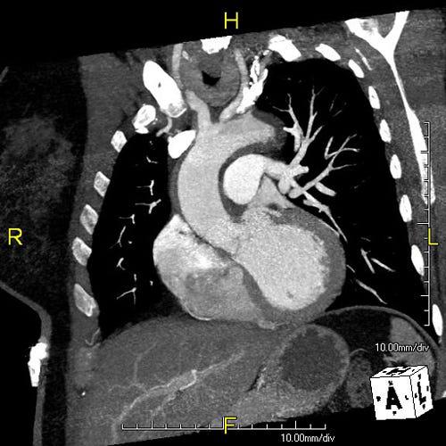
Cardiac MRI provides highly detailed images of the heart’s structures, including the chambers, valves, and blood vessels. It is particularly useful in assessing cardiac function, identifying myocardial infarctions, and evaluating congenital heart defects. Cardiologists may use cardiac MRI for a comprehensive assessment when other imaging techniques may not provide sufficient information.
Nuclear Stress Test:
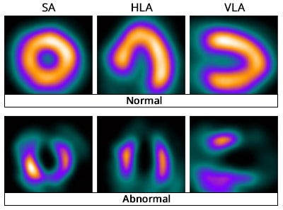
In a nuclear stress test, a small amount of radioactive material is injected into the bloodstream, and a special camera is used to capture images of blood flow to the heart muscle. This test is particularly useful in assessing blood flow during rest and stress, helping identify areas of reduced blood supply and evaluating the extent of coronary artery disease.
Coronary Angiography:
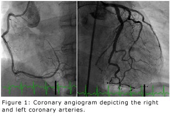
Coronary angiography involves injecting a contrast dye into the coronary arteries and taking X-ray images to visualize blood flow and detect blockages. This invasive procedure is often performed during cardiac catheterization. It is a gold standard for diagnosing coronary artery disease and guiding interventions such as angioplasty and stent placement.
Conclusion
Cardiologists employ a diverse array of diagnostic tests to comprehensively evaluate the heart’s structure and function. From non-invasive procedures like ECG and echocardiograms to more invasive techniques like coronary angiography, each test plays a crucial role in providing valuable insights for accurate diagnosis and effective management of cardiovascular conditions. These tests, when combined and interpreted skillfully by cardiologists, contribute to a holistic understanding of the patient’s cardiac health, allowing for personalized and targeted treatment strategies. Regular cardiovascular assessments with these tests are essential in maintaining heart health and preventing serious complications.